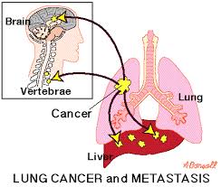
Doctors use various tests to be sure that something seen on an imaging test is really lung cancer, to determine the exact type of lung cancer, and to see how far it may have spread.
A doctor (called a pathologist) who is an expert in using lab tests to diagnose diseases like cancer will look at the cells under a microscope. If you have any questions about your pathology results or any other tests, be sure and ask your doctor. If needed, you can get a second opinion about your report (called a pathology review) by having your tissue sample sent to a pathologist at another lab.
Tests of Tissues and Cells
Sputum cytology. A sample of mucus you cough up from the lungs (phlegm: pronounced "flem") is looked at under a microscope to see if cancer cells are present.
Fine needle biopsy (FNA). A long, thin (fine) needle is used to remove a sample of cells from the area that may be cancer. The sample is looked at in the lab to see if there are cancer cells.
Bronchoscopy. A lighted, flexible tube (called a bronchoscope) is passed through the mouth into the bronchi. This test can help find tumors, or it can be used to take samples of tissue or fluids to see if cancer cells are present. You may be given drugs to make you sleep for this exam.
Endobronchial ultrasound. Ultrasound is a test that uses sound waves to make pictures of parts of your body. For endobronchial ultrasound, a bronchoscope is fitted with an ultrasound device at its tip and is passed down into the windpipe. If areas of concern (such as enlarged lymph nodes) are seen on the ultrasound, a hollow needle can be passed through the bronchoscope and guided by ultrasound into the area to take biopsy samples. The samples are then looked at under a microscope to see if cancer cells are present.
Endoscopic esophageal ultrasound (EUS). This test is much like an endobronchial ultrasound, except an endoscope (a lighted, flexible tube) is used. It is passed down the throat and into the esophagus (the swallowing tube that connects the mouth to the stomach). The esophagus lies just behind the windpipe. This test is done with numbing medicine (local anesthesia) and light sedation.
Ultrasound images taken from inside the esophagus can help find large lymph nodes inside the chest that might contain lung cancer. If areas of concern (such as enlarged lymph nodes) are seen on the ultrasound, a hollow needle can be passed through the endoscope to get biopsy samples of them. The samples are then looked at under a microscope to see if they contain cancer cells.
Mediastinoscopy and mediastinotomy. Both of these tests allow the doctor to look at and sample the structures in the area between the lungs (this area is called the mediastinum) and behind the breast bone. They are done in an operating room while you are in a deep sleep (under general anesthesia). The main difference between is in the place and size of the cut (incision) needed to look into this area.
Thoracentesis and thoracoscopy. These tests are done to check whether fluid around the lungs is caused by lung cancer or by some other medical problem, such as heart failure or an infection. For thoracentesis, the skin is numbed and a needle is placed between the ribs to drain the fluid. The fluid is checked for cancer cells. Thoracoscopy uses a thin, lighted tube connected to a video camera and screen to look at the space between the lungs and the chest wall. By doing this, the doctor can see any cancer deposits on the lung or lining of the chest wall and can take out small pieces of tissue to be looked at under the microscope. Thoracoscopy can also be used to sample lymph nodes and fluid and to tell whether a tumor is growing into nearby tissues or organs.
Bone marrow biopsy: For this test you lie on your side or on your belly. The skin over the back of your hip is cleaned. After the area is numbed, a needle is used to remove a small piece of bone, usually from the back of the hip bone. The sample is checked for cancer cells. This is done mostly to help find if small cell lung cancer has spread to the bones.
Lab Tests and Other Tests
Samples from biopsies or other tests are sent to a lab. There, a doctor looks at the samples under a microscope to find out if they contain cancer and if so, what type of cancer it is. Special tests may be needed to help classify the cancer. Cancers from other organs can spread to the lungs. It's very important to find out where the cancer started, because treatment is different for different types of cancer.
Blood tests: Blood tests are not used to find lung cancer, but they are done to get a sense of a person's overall health. A complete blood count (CBC) shows whether your blood has the correct number of different cell types. This test will be done often if you are treated with chemotherapy because these drugs can affect the blood-forming cells of the bone marrow. Other blood tests can spot problems in different organs such as the kidneys, liver, and bones.
Pulmonary function tests: Pulmonary function tests (PFTs) are often done after a lung cancer has been found. These tests show how well your lungs are working. This is especially important if surgery might be an option in treating the cancer. These tests can give the surgeon an idea of how much lung can be removed or if surgery is a good option at all.


0 nhận xét:
Speak up your mind
Tell us what you're thinking... !